
Yes. Babies with spina bifida can move in the womb and may have normal-looking periods of activity. However, the amount and type of movement can vary based on the form of spina bifida, the location of the spinal opening, and whether the spinal cord or nerves are affected.
Feeling movement—or not feeling much movement—cannot confirm or rule out spina bifida. prenatal ultrasound and, in some cases, fetal MRI are needed to assess the baby’s spine, brain, limbs, and overall development.
For families in the United States, the most useful next step is to discuss movement patterns and scan findings with an obstetrician, maternal-fetal medicine specialist, or genetic counselor.

How spina bifida can affect movement before birth
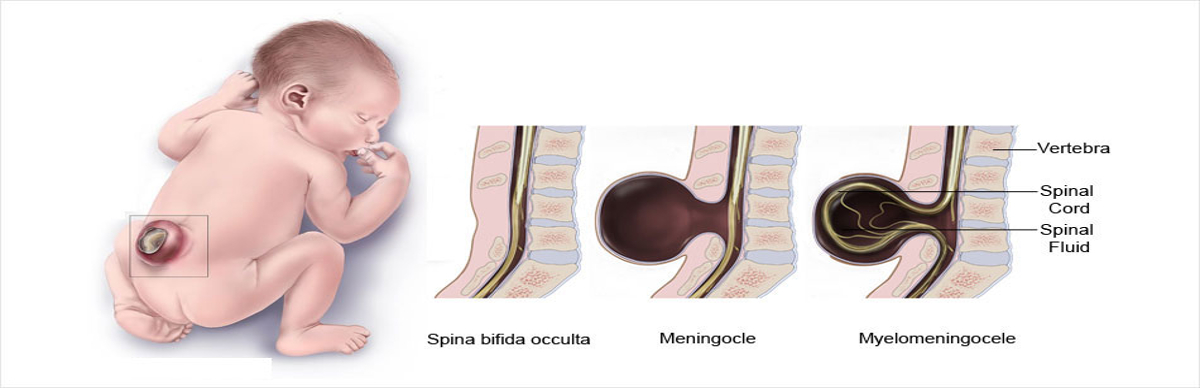
Spina bifida is a neural tube defect. It develops when part of the spinal column does not close fully during early pregnancy. The condition ranges from mild to severe.


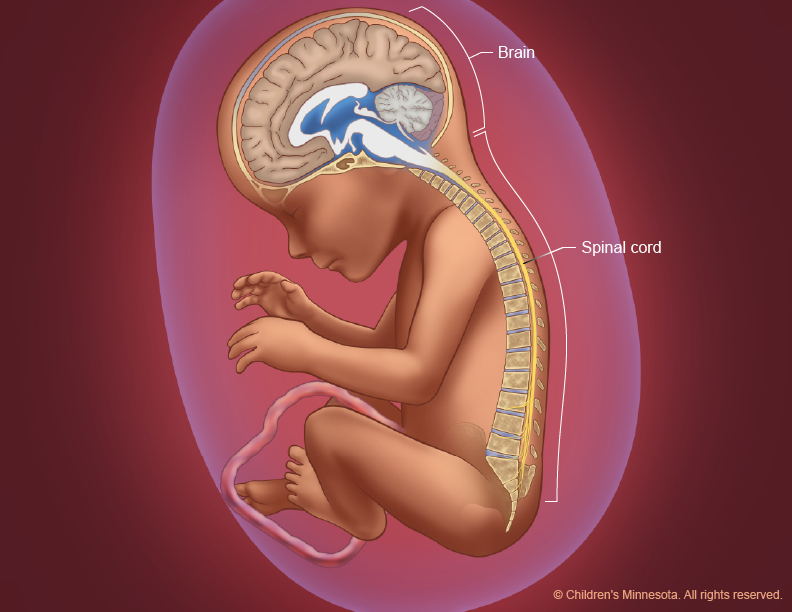
The spinal cord carries signals between the brain and the rest of the body. If the spinal cord or nearby nerves are damaged, a baby may have weakness or reduced movement in parts of the legs. The effect depends mainly on the level and severity of the spinal defect.
Common types of spina bifida
- Spina bifida occulta: A small gap in the bones of the spine. The spinal cord often remains protected, and many people have no symptoms.
- Meningocele: A sac of fluid and spinal coverings pushes through the spinal opening. The spinal cord may not be inside the sac.
- Myelomeningocele: The most severe common form. The sac contains part of the spinal cord and nerves. It is more likely to affect leg movement, sensation, bladder function, or bowel function.
A baby with spina bifida may still move the arms, legs, trunk, or whole body. Movement can be easier to see on an ultrasound than to feel from outside the abdomen.
The key point is that movement does not have to disappear when a baby has spina bifida. A baby may kick, stretch, turn, swallow, open and close the hands, or change position. Some movements may be less strong or less frequent if the spinal nerves are significantly affected.

What fetal movement normally looks like
Fetal movement changes throughout pregnancy. Early movements are often too small for the pregnant person to feel.
Many babies begin making movements during the first trimester. Most pregnant people start to notice movement between about 16 and 22 weeks of pregnancy, although a first pregnancy, the placenta’s position, and the baby’s position can change this timing.
By the third trimester, movement may feel more familiar. It can include:
- Kicks and punches
- Rolling or shifting
- Stretching
- Rhythmic jerks from hiccups
- Pressure in one area of the abdomen
- Changes in movement after eating, resting, or changing position
Movement may feel different as the baby grows. A larger baby has less room for large flips, but movement should not simply stop. Healthy babies also have sleep periods, so activity is not constant.
The American College of Obstetricians and Gynecologists recommends paying attention to a baby’s usual movement pattern. If movement is clearly less than usual, contact the prenatal care team rather than relying only on home methods.

Can ultrasound show whether a baby with spina bifida is moving?
Ultrasound can show fetal movement, but movement alone cannot determine how well the spinal cord is working.


During a detailed ultrasound, the clinician may assess:
- The shape and alignment of the spine
- The location and size of a spinal opening
- Whether a sac is present
- The position of the baby’s legs and feet
- Leg, foot, and body movements
- Brain structures that can be associated with open spina bifida
- Amniotic fluid and overall growth
A standard anatomy scan is commonly performed in the United States around 18 to 22 weeks. If the scan suggests spina bifida or another concern, the obstetric team may recommend a targeted ultrasound with a maternal-fetal medicine specialist.
Ultrasound has limits. A baby may move during the scan even when nerve damage is present. A baby may also be quiet during a short examination for reasons unrelated to spina bifida, such as sleep or position.
In selected cases, fetal MRI may provide more detail about the brain and spine. It does not replace the ultrasound or provide a perfect prediction of future strength, sensation, or mobility.

Does active movement mean the spina bifida is mild?
No. An active baby can still have significant spina bifida.
Fetal movement is only one part of the assessment. A baby with an open spinal defect may move the legs, but the movement may not show how strong the muscles will be after birth. Prenatal imaging also cannot always predict future walking ability.
Doctors consider several findings together:
The level of the spinal defect
A higher defect often has a greater chance of affecting leg function than a lower defect, although individual outcomes vary.Whether the spinal cord is exposed or contained
Myelomeningocele generally carries more risk of nerve-related problems than spina bifida occulta.The appearance of the brain and ventricles
Open spina bifida can be linked with changes such as Chiari II malformation and enlarged brain ventricles.Leg position and movement
The scan may show how the hips, knees, ankles, and feet move, but this is not a complete nerve-function test.Other health findings
The care team may look for additional structural differences or signs that change the treatment plan.
The practical lesson is simple: do not use fetal activity as a test for the severity of spina bifida.
What if the baby seems less active?
A noticeable drop in movement should be treated as a pregnancy concern, whether or not spina bifida is suspected.
If the baby’s movement is less than usual:
- Stop and focus on the baby’s pattern.
- Contact your obstetrician, midwife, or labor and delivery unit for instructions.
- Follow the movement-counting method your care team gave you.
- Seek prompt evaluation if the care team recommends it.
Do not wait until the next scheduled appointment if movement has clearly decreased. Do not rely on a home Doppler to reassure yourself. Hearing a heartbeat does not show that the baby is receiving enough oxygen or that the baby is well.
A quiet period can happen for normal reasons, but reduced movement can also signal a problem with the placenta, fluid, growth, or fetal health. Only an appropriate clinical assessment can sort these possibilities out.


If you are not yet feeling regular movement because the pregnancy is early, lack of movement does not by itself suggest spina bifida. Ask your prenatal clinician when movement should become noticeable for your pregnancy.

What happens after a prenatal spina bifida diagnosis?
A suspected diagnosis usually leads to more detailed counseling and imaging rather than an immediate conclusion about the baby’s future.
A U.S. care team may include:
- A maternal-fetal medicine specialist
- A fetal imaging specialist
- A pediatric neurosurgeon
- A neonatologist
- A pediatric urologist
- A genetic counselor
- A social worker or pregnancy care coordinator
The team may discuss:
- The suspected type and level of spina bifida
- Brain and spine findings
- Possible effects on movement, sensation, bladder, and bowel function
- Delivery planning
- Newborn surgery and hospital care
- Whether fetal surgery is an option
- Benefits, risks, and eligibility requirements for available treatments
Fetal surgery is not appropriate for every pregnancy. Eligibility depends on factors such as the type of lesion, gestational age, maternal health, fetal health, and the requirements of the treatment center. The Management of Myelomeningocele Study found that prenatal repair can reduce some complications in carefully selected pregnancies, but it also carries meaningful risks, including risks to the pregnant patient and the pregnancy.
Ask the team to explain which findings are known, which are uncertain, and what information would change the plan. This is more useful than trying to predict the outcome from movement alone.

Questions to ask your prenatal care team
Consider bringing these questions to an appointment:
- What type of spina bifida does the ultrasound suggest?
- Where is the spinal defect located?
- Is the spinal cord inside the sac?
- Are the baby’s legs and feet moving on the scan?
- What can the scan show, and what can it not predict?
- Are there brain findings such as enlarged ventricles or Chiari II changes?
- Should we have a fetal MRI?
- Do we meet with a pediatric neurosurgeon before delivery?
- Is fetal surgery available for this diagnosis, and what are its risks?
- Where should delivery take place?
- What care might the baby need immediately after birth?
Write down the answers. Prenatal imaging can provide important information, but some questions about strength, sensation, bladder control, and long-term mobility cannot be answered with certainty before birth.

Common misunderstandings about fetal movement and spina bifida
“If the baby kicks, the baby cannot have spina bifida.”
This is false. Babies with spina bifida may move in the womb. Movement does not exclude the condition.
“If movement is weak, the baby definitely has spina bifida.”
This is also false. Reduced movement can have many causes, including fetal sleep, the placenta’s position, pregnancy stage, medication effects, fluid problems, or other medical concerns.
“A normal anatomy scan always rules out spina bifida.”
A detailed ultrasound can detect many cases, especially open spina bifida, but no test detects every condition. Image quality, fetal position, gestational age, and the type of defect matter.
“Prenatal movement predicts whether the child will walk.”
Not reliably. The location and severity of nerve involvement, treatment, muscle strength, joint problems, hydrocephalus, and other factors can affect mobility after birth.
“A home Doppler can replace an examination.”
It cannot. A heartbeat reading does not assess fetal movement, oxygenation, growth, or placental function.
Key takeaways
- Babies with spina bifida can be active in the womb.
- Movement may be normal, reduced, or uneven, depending on the spinal defect.
- Fetal activity cannot diagnose spina bifida or predict future walking ability.
- Ultrasound is the main prenatal test; fetal MRI may add detail in selected cases.
- A clear decrease in your baby’s usual movement needs prompt medical advice.
- Ask a maternal-fetal medicine specialist to explain both the known findings and the limits of prenatal prediction.


Frequently asked questions
Can a baby with spina bifida kick normally?
Yes. Some babies with spina bifida kick and move normally during an ultrasound. Others may show less movement in one or both legs. The pattern depends on the location and severity of nerve involvement.
When can spina bifida be seen on ultrasound?
Many cases can be identified during the detailed anatomy scan, often performed at 18 to 22 weeks in the United States. Earlier scans may detect clues, but timing and visibility vary.
Can spina bifida be missed during pregnancy?
Yes. Small or closed defects can be difficult to see, and no prenatal test detects every case. A suspected finding may require a targeted scan or fetal MRI.
Should I worry if my baby is less active today?
Contact your obstetrician, midwife, or labor and delivery unit if your baby is moving less than usual. Reduced movement should be assessed based on your pregnancy stage and your baby’s established pattern.
Does spina bifida always cause paralysis?
No. Spina bifida has a wide range of effects. Some people have no noticeable symptoms, while others have weakness, paralysis, or problems with bladder and bowel control.
Conclusion
Babies with spina bifida can be active in the womb. Fetal movement may offer a general view of activity, but it cannot show the full health or function of the spinal cord.
If an ultrasound suggests spina bifida, ask for a detailed review with a qualified prenatal care team. If your baby’s usual movement decreases, seek medical advice promptly. The safest answer comes from the full clinical picture—not from kicks alone.
Sources and further reading
- Centers for Disease Control and Prevention: Spina Bifida
- American College of Obstetricians and Gynecologists: Decreased Fetal Movement
- American College of Obstetricians and Gynecologists: Ultrasound Exams
- National Institute of Neurological Disorders and Stroke: Spina Bifida
- NIH: Management of Myelomeningocele Study